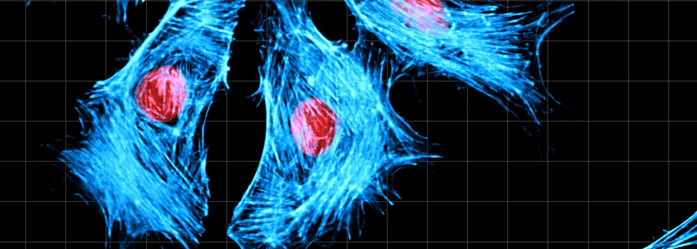
Image analysis is indispensable in scientific research, offering diverse applications for quantitative measurements in cell biology, developmental biology, and neuroscience. Recent advances in fluorescent probes and high-resolution microscopes have enhanced the reliability of biological image processing techniques, profoundly impacting biological sciences research.
An often-overlooked but crucial aspect of cell imaging is quantification and analysis, particularly in contemporary research. Image analysis protocols are increasingly favored by biologists to convert microscopic images into quantifiable measurements. Automated methods help overcome challenges in visually interpreting cell structures and quantifying signals.
Recent developments in artificial intelligence bring new innovative approaches to the field of image processing and analysis.
Various image analysis software packages exist for biological applications, but many are expensive and require high-performance computers. FiJi is considered standard due to its accessibility, platform independence, and applicability for biological researchers.
The ICM.quant platform provides powerful processing and analysis stations to enable the processing and analysis of microscopy images. We are equipped with opensource software like FiJi and commercial software like Imaris, NIS element, Huygens, etc.

