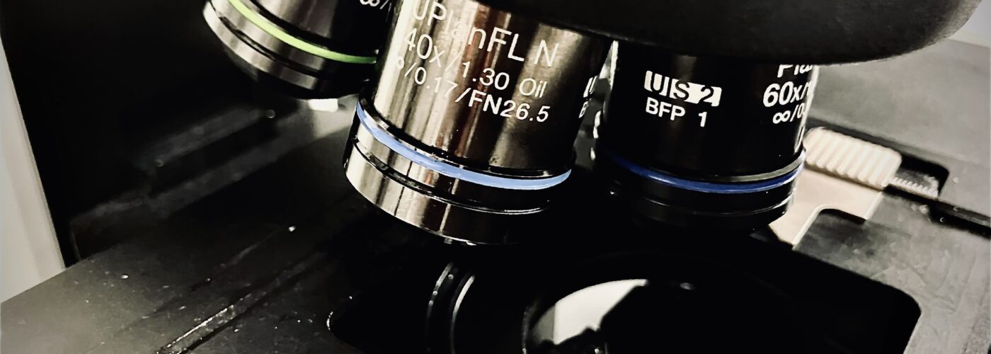
The ICM.Quant platform is equipped with state-of-the-art neuroscience imaging methodologies. The platform offers expertise in photonic microscopy and has a large selection of high-end microscopy imaging methodologies. The platform offers a wide range of services to academic and industrial researchers.
We have a wide variety of equipment:
- Wide-field microscopy: for routine observation of samples in transmitted light or fluorescence.
- Apotome microscopy: to obtain 3D fluorescence images of samples fixed by optical sectioning from a number of images captured with different grid positions.
- Videomicroscopes: for observation of fine living samples in transmitted light or fluorescence.
- Confocal laser scanning microscopy: for 3D fluorescence image acquisition by optical sectioning on medium-thick samples.
- Spinning disk confocal microscopy: enables rapid 3D image acquisition by optical sectioning. This system is suitable for live samples, as it is less photo-toxic than laser scanning confocal microscopy.
- Multi-photon microscopy and intravital microscopy: for in-depth imaging of living or fixed samples.
- Light sheet microscopy on clarified samples: Enables rapid volumetric imaging of small whole organs (mm to a few cm).
- High-content Automated Microscopy: Allows automated acquisition of images in multi-well plates allowing comparative studies before or after treatment.
- STED Super-resolution microscopy: STED (STimulated Emission Depletion) microscopy is a cutting-edge super-resolution technique that transcends traditional diffraction limits in both 2D and 3D dimensions.
Ou network :


